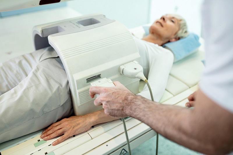
Magnetic Resonance Imaging (MRI)
Magnetic resonance imaging (MRI) produces highly detailed, 3D images of structures inside the body using a magnet and radio waves that allow doctors to diagnose diseases and identify abnormalities.
MRI does not rely on ionizing radiation like an X-ray or CT scan. This is beneficial if someone needs frequent exams to check how a treatment is working or if a condition changes over time.
Because of its magnetic field, MRI may not be an option for people with metal or certain electronic devices in their body – such as pacemakers, aneurysm clips or transdermal patches with metal foil.
How MRIs work
Powerful magnets in the MRI force subatomic particles in the body's tissues called protons to align with the magnetic field. When you add radiofrequency energy to the equation, protons produce signals picked up by a receiver within the scanner to create the images.
An MRI is a painless, non-invasive way to get critical information about your health and concerning conditions.
Conditions evaluated by an MRI
MRI provides more detailed images than CT scans and X-rays. Sometimes, people will have a CT or X-ray and an MRI to get more definitive information about a condition. MRI tests can evaluate soft and hard structures in the body, including bones, tissues, veins, arteries, ligaments, tendons and organs.
MRI scans are used to diagnose:
- Blood flow through the heart and blocked vessels
- Brain abnormalities and tumors
- Breast cancer
- Compression and inflammation of the spinal cord and nerves
- Damage from arthritis
- Damaged cartilage
- Damaged spinal discs
- Heart disease
- Infections
- Joint conditions
- Ligament/tendon tears
- Small stress fractures
- Soft tissue swelling
- Spinal cord abnormalities
- Spinal stenosis
- Torn meniscus
- Tumors
Some MRI scans rely on contrast to highlight the area being studied. The contrast used is not a radioactive material like those used for nuclear medicine imaging. It usually contains the element gadolinium, which increases the speed at which protons align in the magnetic field for sharper images.
Specialists also rely on MRI scans to guide them during procedures, such as an MRI-guided breast or prostate biopsy. Doctors also use a technique called magnetic resonance-guided focused ultrasound to destroy fibroids in the uterus. The MRI guides the doctor to target treatment accurately.
Preparing for an MRI
After your healthcare provider orders the MRI, you will receive a call in about a day to schedule your scan at one of our imaging facilities.
The scheduler will ask you about any metal objects or devices in your body that could pose a threat in the magnetic field of an MRI. This includes:
- Aneurysm clip
- Cochlear implants
- Deep brain stimulators
- Implantable defibrillator
- Insulin pumps
- Pacemakers
- Permanent tattoos or cosmetics
- Shrapnel or bullets
- Transdermal patch with metal foil
- Vagus nerve stimulators
If any of these are present, a radiologist will evaluate and decide if an MRI scan is safe to proceed. Braces do not cause a problem in terms of the magnetic field, but they can distort an image. Materials used for joint replacements are generally safe for an MRI; however, they can distort the image.
In most cases, you will not have to do anything to prepare for an MRI. If you have an abdominal exam, you will be asked not to eat or drink for four hours. We also recommend only a light meal before an MRI if you are having contrast to prevent an upset stomach.

Arriving for an MRI
We ask that you arrive 30 minutes early to register and set up for the exam.
When we call you back, you will put on a gown and place any metal items, handbags, clothes or other items in a locker and take the key with you. You’ll have a chance to use the restroom before the exam.
When it’s time for the MRI, you will lie on a table that slides into the center of a round machine. MRI equipment is loud, so we will provide you with earplugs that protect you from the decibels produced by the MRI. We have additional headphones if you need more protection.
If your exam requires contrast, we will take a series of images without contrast first. We will then administer the contrast either by asking you to drink a solution or through an IV. The contrast works almost immediately.
Some claustrophobic people may have difficulties with an MRI. You may want to talk to your healthcare provider about prescribing a medication you can take before the exam to help with anxiety. If needed, we offer intramuscular sedation administered by one of our highly trained nurses. As a last resort, we can use anesthesia if all other options do not work.
We will also work with our pediatric patients to provide the right environment depending on the child’s needs and reactions.
An MRI can last as little as 10 minutes; however, it usually runs 20-45 minutes. Of course, those with contrast take longer. If you have multiple areas of study, the MRI could take a couple of hours.
Most of the time, you will hear back in about 24 hours about your MRI scan results. Breast MRI results may take 24-48 hours for the radiologist to read the complex and detailed images.
Choosing Spartanburg Regional for an MRI
At Spartanburg Regional, we use the latest techniques and equipment, which our engineers regularly service. Our MRI techs are registered with the American Registry of Magnetic Resonance Imaging Technologists.
Our experienced staff will work hard to put you at ease and help you through what can be a stressful situation.
We have a well-coordinated quality assurance program in which coworkers, supervisors and doctors review MRI studies to ensure we’re using the most effective techniques and best practices when conducting an MRI.


